Tooth Root Pushed Into the Sinus? What Happens Next
Last Updated: September 4, 2026
🕒 12 min read

Written by DMD Alexander K.
Doctor of Dental Medicine, 10+ years of clinical experience, focused on preventive dentistry and patient education. Learn more on the About page.

Table of Contents
Wait, can a tooth root actually end up in my sinus? 🦷
Yes. Genuinely, anatomically, documented-since-forever yes. Not urban legend, not a scare story a nervous friend told you before your wisdom tooth appointment — a real, recognized thing that happens during upper back tooth extractions, occasionally on purpose from the dentist’s perspective and occasionally very much not.
Here’s the sentence that should calm you down before anything else scares you: most of the time, this gets caught immediately, right there in the chair, and gets handled before you even leave the office. [1,2] The unsettling version — the one this article spends real time on — is the smaller share of cases where nobody notices for months, sometimes years, and a root fragment just sits there, quietly filing a change-of-address form with your sinus.
🦷 This article is part of our Adult Oral Health Guide Adult Oral Health Guide, where we break down the most common dental problems and how to actually deal with them.
Related Reads 🔗
How to Know If a Dentist Is Good: 10 Trustworthy Signs
Not sure if your dentist is any good? Here are 10 trustworthy signs that help patients choose the right dentist — and avoid the ones they shouldn’t trust.
Gum Turned White After a Dental Injection? What It Means
A pale white patch right after the needle isn't the same as a sore that shows up days later. Here's the difference — and when to actually worry.

Bruised Cheek After a Dental Injection? What It Means
A dental injection isn't supposed to leave a bruise — but sometimes it does. Here's why, how long it lasts, and when it's actually a problem.
What’s actually happening (why upper back teeth are the risk) 🔬
Your upper back teeth — specifically the molars, and to a lesser degree the premolars — have roots that sit remarkably close to the floor of your maxillary sinus. In some people, “close” undersells it: the only thing separating a root tip from open sinus space is a thin membrane, no bone at all. [1,2] Nature, apparently, was not consulted on the convenience of this arrangement for anyone holding a dental elevator.
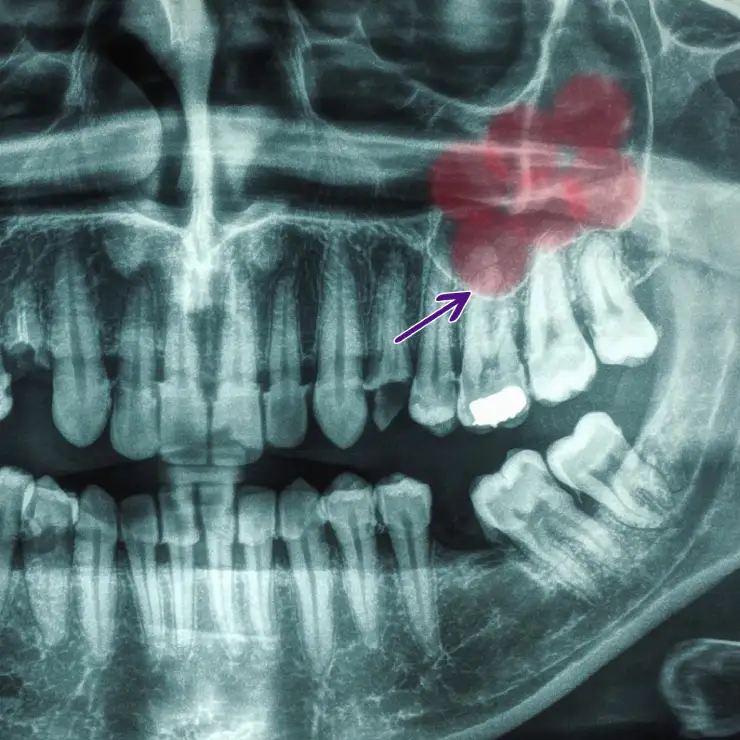
Red area highlights the sinus chamber and the arrow shows the border of the maxillary sinus. There you can see the roots of the upper molars in close proximity to the sinus floor. Even dough it looks like the roots are in the sinus, that doesn't mean they actually are, it like when you overlap two objects in a 2D image — one can appear in front of the other without actually being inside it. The dentist and his experience help him interpret these images correctly, and prepare up front for any potential complications.
The usual sequence of events, when this goes wrong: a root fractures during extraction — not uncommon on its own — and instead of coming out cleanly, a fragment gets pushed the wrong direction. Straight up, straight into the sinus. This is almost always the result of blind instrumentation (which often occurs and sometimes is unavoidable) — an elevator applying force apically without a clear view of exactly where that force is sending the fragment. [2]
One tooth is disproportionately responsible. The palatal root of the upper first molar accounts for close to 80% of all displaced roots. [2,7] The second molar picks up most of the remainder, at around 20%. [2,7] Premolars and canines get displaced roots too, technically, but rarely enough that they’re barely worth mentioning here — this is overwhelmingly a first-molar problem, specifically its palatal root, specifically because that root sits furthest from the operator’s direct line of sight and closest to the sinus floor. [7]
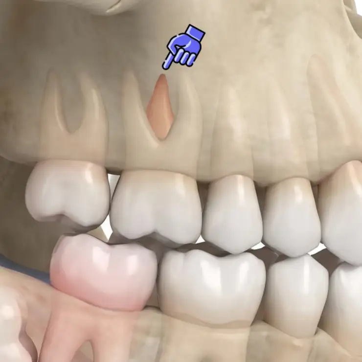
The palatal root goes deep into the bone and sits closest to the sinus floor, making it particularly susceptible to displacement into the sinus during extraction.
What actually raises your personal risk: an especially large, air-filled sinus — which tends to expand with age or after nearby teeth have been missing for a while — a genuinely difficult extraction, and, bluntly, excessive apical force from an elevator that isn’t being used with restraint. [1,2,8] None of these are things you can control from the patient chair. They’re either your anatomy or your dentist’s technique, and only one of those two is something worth vetting in advance.
Once a fragment is up there, it lands in one of three spots: floating loose inside the sinus itself after punching through the membrane, wedged between the bone and the membrane without fully breaching it, or — the least dramatic outcome — trapped under the tissue just outside the sinus proper without ever actually getting in. [1,2,7] Telling these apart from an X-ray alone is genuinely tricky, which is part of why this condition has a habit of getting missed.
How it’s usually caught — and how it sometimes isn’t 🔍

This is a good quick test for detecting if there is a communication between your mouth and your sinus. Hence, if it's negative, it doesn't exclude the possibility entirely.
The obvious version, caught in real time: the root simply vanishes mid-procedure — one second it’s there, the next it isn’t — often paired with a very specific, very unpleasant sign: bubbles appearing in the socket when you rinse, because air and fluid are now moving between your mouth and your sinus through a hole that wasn’t there ten minutes ago. [1,2] Some patients even notice fluid escaping from their nose, usually right on the first rinse after the procedure. A post-op X-ray confirms it: the root that was supposed to be in a kidney dish is now, somehow, up near the sinus floor. [1,2]
Getting the imaging right matters more than you’d think. A standard periapical X-ray can show the root sitting higher than it should, but the flat, single-plane panoramic X-ray many offices default to can genuinely miss it entirely if the root doesn’t happen to land in the exact slice the machine captured. [2] A CT or cone-beam CT is the actual gold standard here — full three-dimensional placement, no guessing. [1,3,5] If there’s any doubt after a routine film, that’s the next step, not a shrug.
The version that slips through: sometimes nobody notices at the time. No dramatic disappearance, no bubbling, a socket that looks unremarkable, and a fragment quietly sitting in a spot small enough or positioned well enough that it doesn’t announce itself. This is precisely the scenario the rest of this article exists for.
What it can feel like months or years later 📅
This is the section most other content on this topic skips entirely, because most of what’s written about this is aimed at dentists holding an elevator, not at someone quietly wondering whether their sinus problems have an origin story from two years ago.
Here’s the honest range: a small, well-tolerated fragment can sit there indefinitely and cause nothing at all. Research examining these roots under a microscope found some become genuinely incorporated — coated in reparative tissue, surrounded by sinus lining that shows barely any inflammation, functionally adopted by the body rather than rejected. [6] This tends to happen when the root wasn’t infected going in, the fragment is small, and it landed submucosally rather than fully loose inside the sinus cavity. [6] Dentists sometimes describe this as putting the root “on probation” — left in place, watched over time, causing no trouble.
The less lucky version looks like this instead: a sinus infection that doesn’t behave like your usual sinus infection — persistent, one-sided, oddly resistant to whatever your doctor’s first-line treatment was, because there’s a literal foreign object sitting in there that antibiotics alone were never going to resolve. Sometimes it’s a lingering bad taste or smell that won’t go away. Sometimes it’s chronic nasal congestion on just one side, which is a genuinely useful clue precisely because most ordinary sinus issues don’t respect that kind of anatomical boundary. Occasionally, it’s simply a persistent connection between your mouth and your sinus — air moving where it shouldn’t, fluid coming through your nose when you drink, a whistling sound you’ve started associating with a specific tooth gap. [1,7]
If any of that sounds like something you’ve been living with since a molar extraction that happened longer ago than you’d like to admit — that’s worth mentioning to a dentist specifically, by name, rather than continuing to treat it as an unrelated, ordinary sinus problem.
How it’s treated 🛠️
If it’s caught immediately, in the chair: your dentist sits you upright — gravity matters here, and lying flat makes a stray fragment more likely to migrate further back — confirms the location on imaging, and attempts gentle suction directly through the socket. [7] If that doesn’t retrieve it, irrigating with sterile saline and suctioning again is the next move. [7] What should not happen at this point is repeated blind poking around with an instrument, hoping to get lucky — that risks pushing the fragment even further in. [1,2,7] If simple suction doesn’t work, the right move is a referral to an oral and maxillofacial surgeon, not persistence. [1,2,7]
If it’s small, uninfected, and well-positioned: sometimes the actual treatment is patience. Genuinely small fragments in a healthy sinus can clear out on their own over days to weeks, carried along by your sinus’s own natural mucus-clearing mechanism toward the drainage opening and eventually out through your nose. [5] This isn’t guesswork — it gets confirmed with follow-up imaging showing the fragment actually moving in the right direction, not just hoped for. [5]
If it needs surgical retrieval — because it’s larger, infected, or conservative monitoring failed — there’s a genuine menu of options, and which one gets used depends heavily on timing and location:
- Through the original socket, if the extraction is recent enough that it hasn’t fully healed — minimally invasive, done under local anesthesia, using a thin suction catheter guided by imaging to find and capture the fragment. [10]
- The Caldwell-Luc approach, involving a small bony window created near the canine area to get direct visual access to the sinus — the traditional workhorse for roots sitting high up or after the socket has already closed. [1,3,8]
- Endoscopic retrieval, increasingly the preferred modern option — a small camera, excellent visualization, smaller incisions, and meaningfully less collateral damage to nearby structures than the older open approaches. [1,8]
None of these are exotic. All of them are established, documented procedures with predictable recovery expectations, performed by oral and maxillofacial surgeons who have, statistically, seen this exact situation before.
What not to do if this just happened to you 🚫
Don’t blow your nose. Forcefully, gently, at all, for the time being. Increased pressure is exactly what pushes fluid and infection in the wrong direction through a fresh communication. [7]
Don’t skip the follow-up because you feel fine. Feeling fine today doesn’t rule out a slow-brewing infection or a communication that hasn’t closed properly yet.
Don’t let anyone talk you into “just waiting and seeing” without imaging first. Watchful waiting is a legitimate strategy — but only when it’s an informed one, based on a confirmed small, uninfected, well-positioned fragment, not a guess made without ever actually looking. [5,6]
Don’t panic-research your way into assuming the worst. The genuinely dangerous complications — infections spreading toward the brain, that kind of territory — exist in the literature specifically because they’re rare enough to be worth writing up individually. [1] That’s the zebra case, not the typical outcome, and it’s mentioned here so you know it exists, not so you spiral over it.
When to actually see someone 📞
Get evaluated, and don’t wait it out, if:
- You notice bubbling, fluid, or a strange taste when rinsing shortly after an upper back tooth extraction — that’s the immediate, obvious sign something’s off. [1,2]
- You’ve had one-sided sinus symptoms — congestion, pressure, drainage, or a persistent bad smell or taste — that started sometime after an upper molar extraction and never fully resolved, even if that extraction was a while ago.
- A “sinus infection” keeps not responding to whatever your regular doctor’s standard treatment is, especially if it keeps coming back in the same spot.
- You’re mid-extraction and your dentist mentions a root has gone missing — that’s their moment to refer you onward if simple retrieval doesn’t work immediately, and it’s a completely reasonable question to ask about directly.
Upper molar extractions, given how close that anatomy runs, genuinely benefit from an experienced hand — choosing the right dentist matters here in a very literal, anatomical way, not just a general “good bedside manner” way.
Bottom line 🎯
A tooth root ending up in your sinus during an upper back tooth extraction is a genuine, documented anatomical risk — not a horror story, not a freak accident, and, in the majority of cases, something caught and handled the same day it happens. [1,2] The part worth taking seriously is the smaller, quieter possibility: a fragment that goes unnoticed, sits for months or years, and eventually announces itself as a stubborn, one-sided sinus problem that doesn’t behave like an ordinary one.
This is not, by default, a sign anyone did anything wrong. The anatomy in that specific region is simply unforgiving — sometimes there’s no bone at all between a root tip and open sinus space, and even careful, competent extractions carry a real, acknowledged risk here. [1,2] What actually matters is prompt recognition when it happens, appropriate imaging when there’s any doubt, and treatment matched to the size and behavior of the fragment rather than a blind guess either way.
Sources
- [1] Beech AN, Farrier JN. The importance of prompt referral when tooth roots are displaced into the maxillary antrum. *Dental Update*. 2016;43(8):760-765. DOI: 10.12968/denu.2016.43.8.760
- [2] Barclay JK. Root in the maxillary sinus. *Oral Surgery, Oral Medicine, Oral Pathology*. 1987;64(2):162-164. DOI: 10.1016/0030-4220(87)90083-1
- [3] Asmael HM. The Modified Caldwell-Luc Approach in Retrieval of Accidentally Displaced Root into the Maxillary Sinus. *Journal of Craniofacial Surgery*. 2018;29(2):e130-e131. DOI: 10.1097/scs.0000000000004189
- [4] Sun R, Zheng LW, Sun YQ, Li TT, Ren JG, Zhao JH. Is it safe and effective to extract impacted maxillary tooth adjacent to maxillary sinus via submaxillary sinus membrane space approach?—a randomized controlled trial. *Clinical Oral Investigations*. 2023;27(10):6081-6087. DOI: 10.1007/s00784-023-05223-8
- [5] Tocaciu S, Sillifant P. Spontaneous clearance of a dislodged root in the maxillary antrum. *Oral Surgery*. 2018;11(3):224-227. DOI: 10.1111/ors.12326
- [6] Herd JR. The tooth root within the maxillary sinus. *Australian Dental Journal*. 1974;19(2):77-83. DOI: 10.1111/j.1834-7819.1974.tb04306.x
- [7] Laskin DM. Management of Oroantral Fistula and other Sinus-Related Complications. *Oral and Maxillofacial Surgery Clinics of North America*. 1999;11(1):155-164. DOI: 10.1016/s1042-3699(20)30943-2
- [8] Wang X, Li S, Huang Z, Cai Q, Huang Z, Chen W. Removal of the Residual Roots Displaced Into the Maxillary Sinus via Endoscopy. *Journal of Craniofacial Surgery*. 2018;29(4):1034-1036. DOI: 10.1097/scs.0000000000004382
- [9] Cheng L, Papesch M. Removal of dislodged roots from maxillary antrum using minimal access sinuscopy and eicken antrum cannula. *International Journal of Oral and Maxillofacial Surgery*. 2019;48:288. DOI: 10.1016/j.ijom.2019.03.880
- [10] Cheng H, Sproat C, Patel N. A novel transalveolar approach for retrieval of displaced tooth roots from the maxillary sinus using a modified suction catheter: technical note. *Oral Surgery*. 2019;13(1):12-14. DOI: 10.1111/ors.12444
FAQ: Tooth Root Displaced Into the Sinus Real Answers❓
What happens if a tooth root ends up in the sinus during an extraction?
How is a displaced root in the sinus usually caught?
Can this go unnoticed for years?
What symptoms should make me suspect this happened to me?
How is a root fragment in the sinus treated?
What should I avoid doing if this just happened during my own extraction?
How long does recovery from treatment take?
Is this a sign my dentist did something wrong?
About the Author: DMD Alexander K.
Doctor of Dental Medicine with clinical experience treating adults and children. This site focuses on practical prevention, symptom education, and helping patients make informed decisions.
Learn more on the About page.